

Below, you can find some photos of our common equipment for processing and characterization.of Advanced Materials and Systems for Energy laboratories:
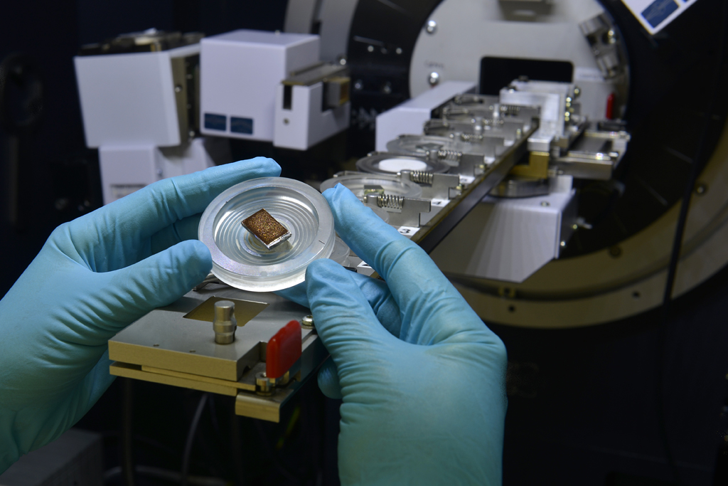
X-ray powder diffraction (XRD) is a powerful non-destructive analytical technique applied for identification of crystalline phases and its quantification and that also provides detailed crystallographic structure information such as crystal structure, % crystallinity, unit cell, crystallite size, strain, preferred orientation and layer thickness for a wide range of materials from powders to solids, thin films and nanomaterials.
Instrumentation: XRD D8 Advance (Bruker)
Accessory: Reactor chamber, XRK 900 (Paar Anton) for in-situ X-ray diffraction experiments from room temperature to 900ºC.
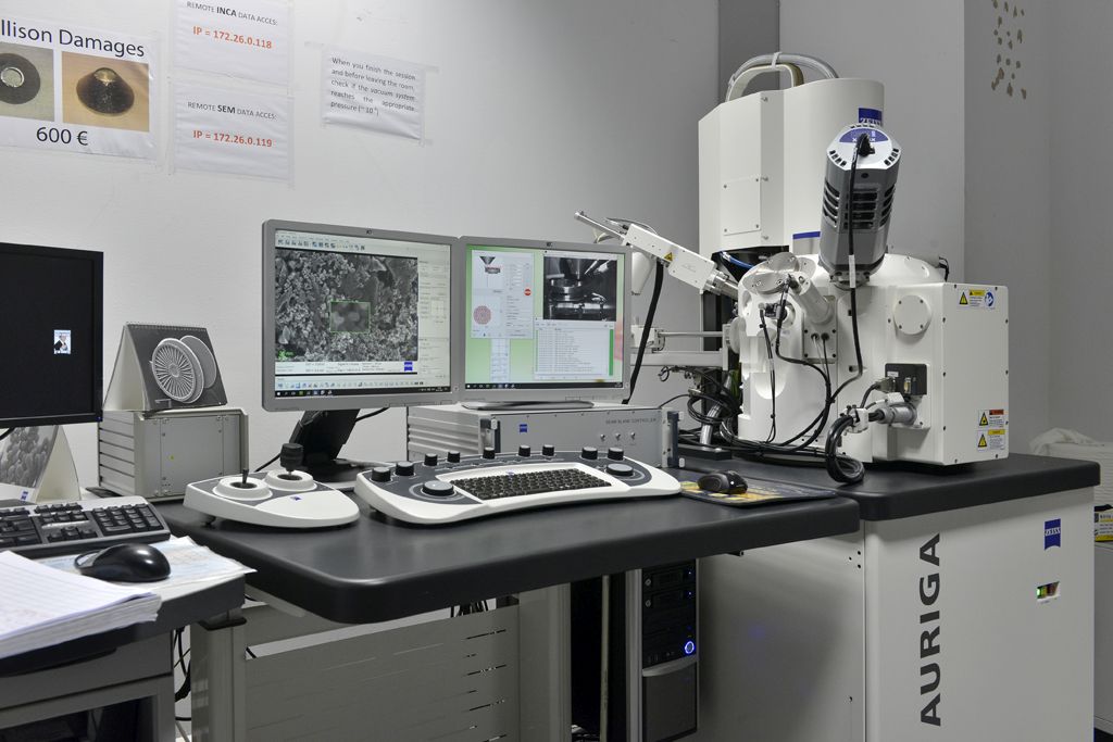
The Field Emission Scanning Electron Microscope (FE-SEM) is a microscope that provides high resolution and high depth of field images of the sample surface. The SEM uses a focused high-energy beam of electrons scanning over the area of interest of the samples to produce images. The electrons in the beam interact with the atoms of the sample generating a variety of signals that carry different information about the sample. The most commonly signals detected are: the secondary electrons (SE) which give topographic and morphological images, the backscattered electrons (BSE) illustrate contrast in chemical composition, the X-ray provide elemental identification and quantitative compositional information with the Energy Dispersive X-ray spectroscopy detector. Instrumentation: FE-SEM serie Auriga 60 (Carl Zeiss) coupled with Oxford INCA Instruments Energy Dispersive Spectrometer (EDS)
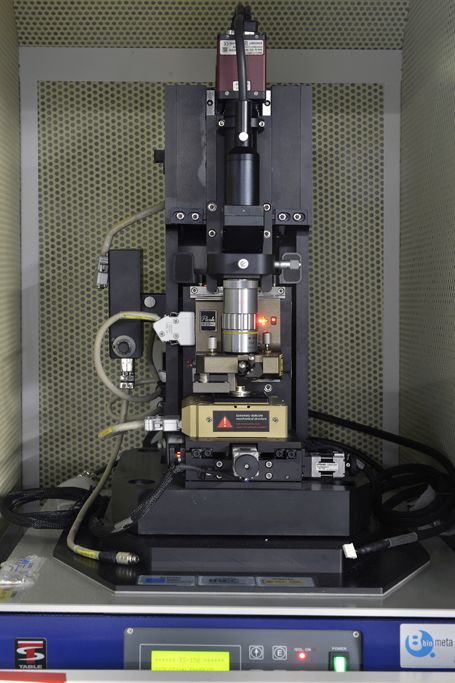
Atomic Force Microscope (AFM) is a surface scanning instrument that provides 3D surface topographic maps in a sub-nanometer scale resolution by probing with a very sharp tip which vertical movements are recorded and used to create the image. It can also be used to probe other surface properties such as : mechanical, electrical, thermal.
Instrumentation: AFM XE-100 (Parks Systems)
Accessories: SthM . Scanning Thermal microscopy
CP-AFM : Conductive AFM
Universal Liquid Cell with cooling stage
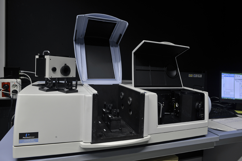
UV/VIS/NIR spectroscopy is an analytical technique to determine the optical properties (transmittance, reflectance and absorbance) of liquids (Standard module), powders (Praying Mantis module) and solids (Integrating Sphere module) in the 180-3300nm wavelength range.
Instrumentation: Lambda 950 UV-Vis-NIR Spectrophotometer (Perkin Elmer)
Accessories: Integrating Sphere of 150mm
Praying Mantis
Transmission Electron Microscope (TEM) is a microscope technique in which a high energy electrons beam (e.g 80 to 200 kV) is transmitted through an ultra thin specimen. The interactions between the electrons and the sample provide images with up to atomic resolution if the beam energy is high enough. Further information can be obtained from the diffraction of electrons by the crystallographic lattice.
Instrumentation: TEM Libra 120 (Carl Zeiss)
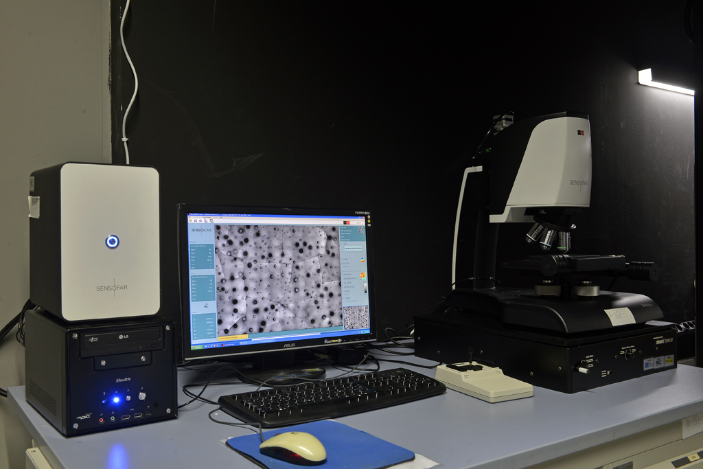
Confocal microscopy is a type of optical microscopy in which most of the out-of-focus signal is blocked by a pinhole screen and an image corresponding to only a thin section of the sample is acquired. By scanning the sample in the Z-axis and combining the 2D acquired images with the Z position information, it is possible to reconstruct the 3D surface morphology (topography) of the sample.
Instrumentation: PLux neox (Sensofar)